Tricuspid Stenosis - Moderate
Virtual Auscultation
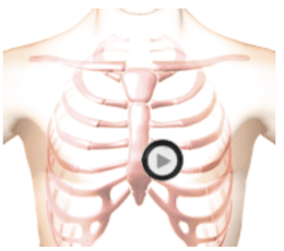

The patient's position is supine.
Lesson
This is an example of moderate tricuspid stenosis which is most commonly due to rheumatic heart disease. The first heart sound is increased in intensity due to moderate thickening of the tricuspid valve leaflets. The second heart sound is normal and unsplit. Systole is silent. There is a tricuspid opening snap followed by a diamond shaped low frequency murmur. Use the bell of the stethoscope to hear this murmur. There is a second murmur in late diastole due to contraction of the right atrium. In the animation you can see the turbulent blood flow from the right atrium into the right ventricle. You can see the moderately thickened tricuspid valve leaflets and the moderately enlarged right atrium. The excursion of the tricuspid valve leaflets is moderately decreased. For this condition the murmur intensity and tricuspid opening snap increase with inspiration.Waveform
Heart Sounds Video
In the cardiac animation take note of the turbulent blood flow from the right atrium into the right ventricle. Observe the moderately thickened tricuspid valve leaflets and the moderately enlarged right atrium. The excursion of the tricuspid valve leaflets is moderately decreased.
For this condition the murmur intensity and tricuspid opening snap increase with inspiration.
Authors and Sources
Authors and Reviewers
-
Heart sounds by Dr. Jonathan Keroes, MD and David Lieberman, Developer, Virtual Cardiac Patient.
- Lung sounds by Diane Wrigley, PA
- Respiratory cases: William French
-
David Lieberman, Audio Engineering
-
Heart sounds mentorship by W. Proctor Harvey, MD
- Special thanks for the medical mentorship of Dr. Raymond Murphy
- Reviewed by Dr. Barbara Erickson, PhD, RN, CCRN.
-
Last Update: 12/11/2022
Sources
-
Heart and Lung Sounds Reference Library
Diane S. Wrigley
Publisher: PESI -
Impact Patient Care: Key Physical Assessment Strategies and the Underlying Pathophysiology
Diane S Wrigley & Rosale Lobo - Practical Clinical Skills: Lung Sounds
- Essential Lung Sounds
Diane S. Wrigley, PA-C
Published by MedEdu LLC - PESI Faculty - Diane S Wrigley
-
Case Profiles in Respiratory Care 3rd Ed, 2019
William A.French
Published by Delmar Cengage - Essential Lung Sounds
by William A. French
Published by Cengage Learning, 2011 - Understanding Lung Sounds
Steven Lehrer, MD
- Clinical Heart Disease
W Proctor Harvey, MD
Clinical Heart Disease
Laennec Publishing; 1st edition (January 1, 2009)