Mitral Stenosis Severe and Regurgitation Mild - Rheumatic Origin
Virtual Auscultation
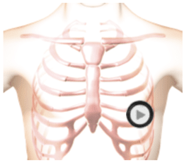

The patient's position is supine left side down.
Lesson
This is an example of severe mitral stenosis combined with mild mitral regurgitation in a patient with rheumatic heart disease. The first heart sound is slightly louder than normal. The second heart sound is unsplit. There is an opening snap fifty milliseconds after the second heart sound. There is a low-frequency murmur filling all of diastole. The first two-thirds of the murmur is diamond shaped and the last third is a crescendo. There is a rectangular medium frequency murmur which fills the first half of systole. In the anatomy video you can see an enlarged left atrium and thickened mitral valve leaflets which barely moved. The turbulent blood flow represents the systolic and diastolic murmurs.Waveform
Heart Sounds Video
Review the animation. Notice an enlarged left atrium and thickened mitral valve leaflets which barely moved. The turbulent blood flow represents the systolic and diastolic murmurs.
Authors and Sources
Authors and Reviewers
-
Heart sounds by Dr. Jonathan Keroes, MD and David Lieberman, Developer, Virtual Cardiac Patient.
- Lung sounds by Diane Wrigley, PA
- Respiratory cases: William French
-
David Lieberman, Audio Engineering
-
Heart sounds mentorship by W. Proctor Harvey, MD
- Special thanks for the medical mentorship of Dr. Raymond Murphy
- Reviewed by Dr. Barbara Erickson, PhD, RN, CCRN.
-
Last Update: 12/11/2022
Sources
-
Heart and Lung Sounds Reference Library
Diane S. Wrigley
Publisher: PESI -
Impact Patient Care: Key Physical Assessment Strategies and the Underlying Pathophysiology
Diane S Wrigley & Rosale Lobo - Practical Clinical Skills: Lung Sounds
- Essential Lung Sounds
Diane S. Wrigley, PA-C
Published by MedEdu LLC - PESI Faculty - Diane S Wrigley
-
Case Profiles in Respiratory Care 3rd Ed, 2019
William A.French
Published by Delmar Cengage - Essential Lung Sounds
by William A. French
Published by Cengage Learning, 2011 - Understanding Lung Sounds
Steven Lehrer, MD
- Clinical Heart Disease
W Proctor Harvey, MD
Clinical Heart Disease
Laennec Publishing; 1st edition (January 1, 2009)