Hypertrophic Cardiomyopathy - Auscultation Lesson
Hypertrophic cardiomyopathy associated murmur is an early peaking, harsh, diamond shaped, systolic murmur. It begins at the onset of systole and stops well before S2. A fourth heart sound gallop is also present in diastole. View the waveform to see these features.
Observe that S1 intensity has increased due to a hyperdynamic left ventricle. S2 is single.
On the anatomy video, observe that the contraction of the left ventricle is strong and occurs in a reduced amount of time. Anatomically, the septal wall is very much thicker than the rest of the ventricle, but this is not shown in the animation.
The strong contraction of the left ventricle causes the anterior leaflet to be sucked into the ventricle, blocking the flow into the aorta and causing an aortic murmur. At the same time turbulent flow from the left ventricle to the left atrium causes a second murmur. Since the two murmurs occur at the same time, you hear a single murmur.
You can hear the difference between the two murmurs by moving the stethoscope head the aortic to the mitral valve area. First, you will hear the diamond shaped aortic murmur and later the rectangular pansystolic murmur.
Listen
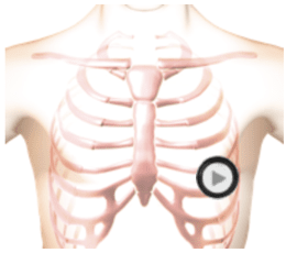
The patient's position is supine.